
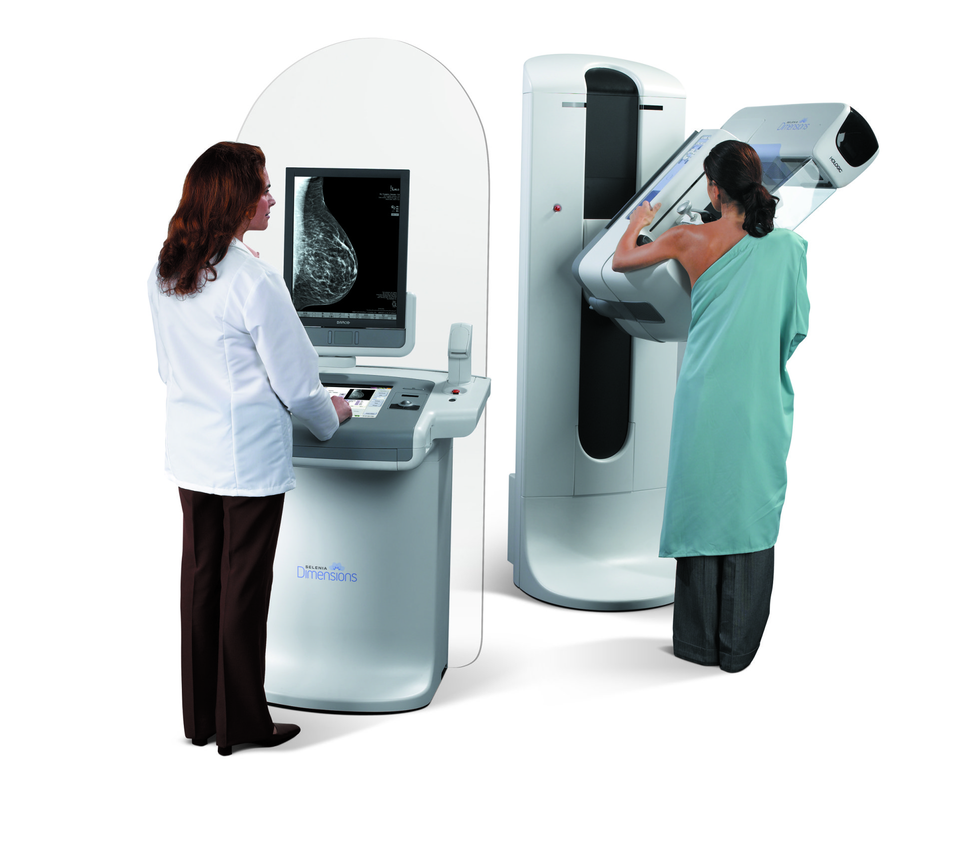
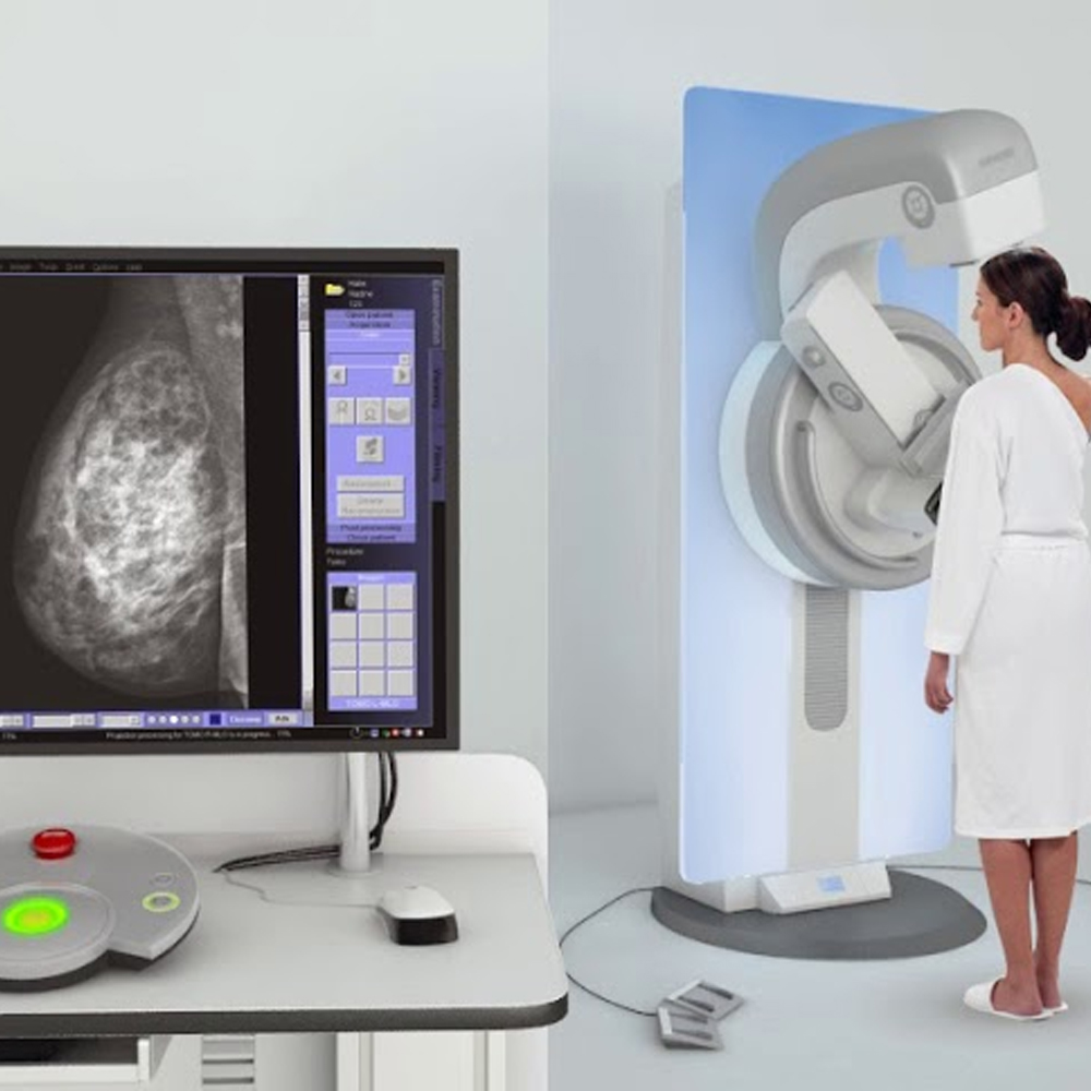
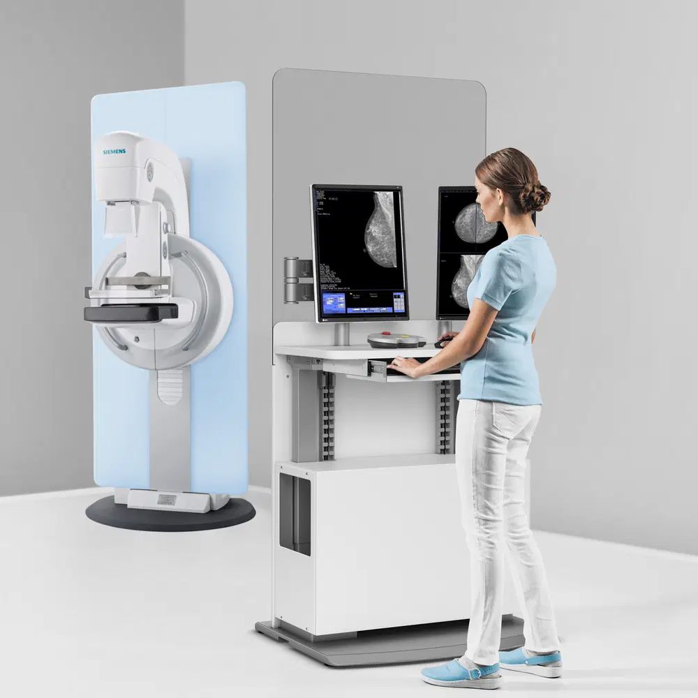
Breast tomosynthesis is a more advanced way to view the breast for any changes, such as signs of breast cancer. Like a traditional mammogram, it uses low-dose X-rays to take pictures of the breast. Breast tomosynthesis captures many images from different angles as the X-ray machine moves around the breast in a small arc.
These images are then combined by a computer to create a detailed, 3D view of the breast tissue. This allows doctors to see the tissue in thin “slices,” much like flipping through the pages of a book. This technique helps radiologists, who are doctors trained to interpret screening images, to spot any abnormal areas that might be hidden on a traditional 2D mammogram.
The 3D images make it easier to detect small tumors and other changes, especially in women with dense breast tissue. While breast tomosynthesis uses a very low dose of X-rays, it is often performed alongside a standard 2D mammogram, so the overall radiation exposure is slightly higher. Despite this, the benefits of more accurate detection and fewer follow-up tests may outweigh the risks.


There are no reviews yet.